RA Imaging: What It Is, How It Helps, and What Doctors Look For
When you have rheumatoid arthritis, a chronic autoimmune condition that attacks the joints, causing pain, swelling, and long-term damage. Also known as autoimmune arthritis, it doesn’t just make you ache—it quietly eats away at bone and cartilage over time. That’s where RA imaging, a set of medical scans used to see inside joints and track how the disease is changing. Also known as arthritis imaging, it’s not just a picture—it’s a timeline of damage. Without it, doctors are guessing. With it, they can tell if your treatment is working, if the damage is speeding up, or if you need to switch drugs before your joints are ruined.
RA imaging includes X-rays, the oldest but still essential tool that shows bone erosion and joint space narrowing, MRI, a high-res scan that catches inflammation and early bone damage before X-rays can, and ultrasound, a real-time, radiation-free method that sees swollen tendons and fluid in joints. Each one tells a different part of the story. X-rays show what’s already broken. MRI shows what’s about to break. Ultrasound shows what’s still active and inflamed. Together, they help doctors decide: Is this drug working? Should we go stronger? Is it time for a biologic?
These scans aren’t just for diagnosis—they’re for tracking. A single X-ray might look okay, but comparing it to one from a year ago can reveal new holes in your bones. An MRI might show swelling in your wrist that you didn’t even feel. Ultrasound can spot fluid building up in your knuckles before your hand swells up. This isn’t science fiction—it’s routine care for people with RA. And it’s why some patients stay mobile for decades while others lose function quickly: it comes down to how often and how well their damage is being seen.
What you’ll find in the posts below isn’t theory. It’s real advice from people who’ve been through it—how to prepare for scans, what the results actually mean, why your doctor ordered one scan over another, and how imaging changes treatment. You’ll learn what to ask when you get your report, why some insurance companies push back on MRIs, and how early detection can save your hands, knees, and quality of life. This isn’t about fancy tech—it’s about catching damage before it catches you.
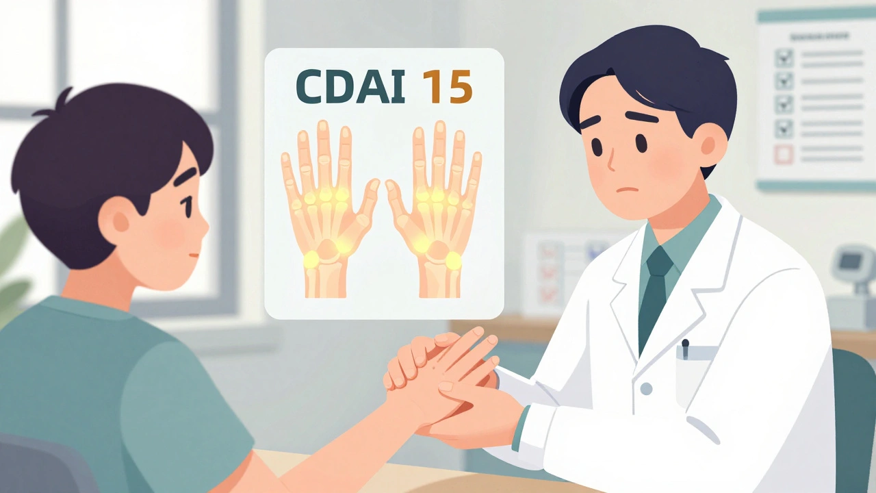
CDAI, DAS28, and imaging are key tools for tracking rheumatoid arthritis. Learn how each works, when to use them, and why combining them leads to better outcomes and less joint damage.